Hip And Leg Bone Diagram : The Hip Joint Articulations Movements Teachmeanatomy / He leg's main function in the human is for locomotion and support of the rest leg bones, learn what and where these are as well as their functions and how we use them.
Hip And Leg Bone Diagram : The Hip Joint Articulations Movements Teachmeanatomy / He leg's main function in the human is for locomotion and support of the rest leg bones, learn what and where these are as well as their functions and how we use them.. The foot bones shown in this diagram are the talus, navicular, cuneiform, cuboid, metatarsals and calcaneus. Download this free vector about diagram showing the hip bone treatment, and discover more than 13 million professional graphic resources on freepik. This lengthy bone connects with the knee at one finish and the ankle on the different. The main reason for its in depth use is that they are corrosion resistant and can be employed in any type of surroundings. Bones are mostly made of the protein collagen , which forms a soft framework.
The bones of the leg are the femur, tibia, fibula and patella. Skeletal hand diagram just another wiring diagram blog. Bones have an internal structure similar to a honeycomb, which makes. Right hip bone in situ & ex situ oriented obliquely to face the hip joint socket (acetabulum). Want to learn more about it?
Hip Muscles The Definitive Guide Biology Dictionary from biologydictionary.net Tensor fascia lata trigger point in it band and hip pain dr perry details the tensor fascia late trigger point that cause hip pain and it band syndrome hip injuries hip disorders take a look at some mon and not so. The foot bones shown in this diagram are the talus, navicular, cuneiform, cuboid, metatarsals and calcaneus. Learn more about the anatomy of the hip using these hip diagrams that will show you the detailed hip joint anatomy hip bones ligaments muscles. The transverse ligaments surround the hip the hip abductors are acting normally tilting the pelvis upwards when the opposite leg is raised. Diagram table of the 1 hip bone showing mineral the. Bones of the hip joint. Previously covered was the hip and we shall now cover the femur (upper leg), patella (kneecap) and the tibia and fibula (the two lower leg elements). The bones of the leg are the femur, tibia, fibula and patella.
When the leg is stretched out, the knee joint is placed on a straight line with the hip and ankle (left).
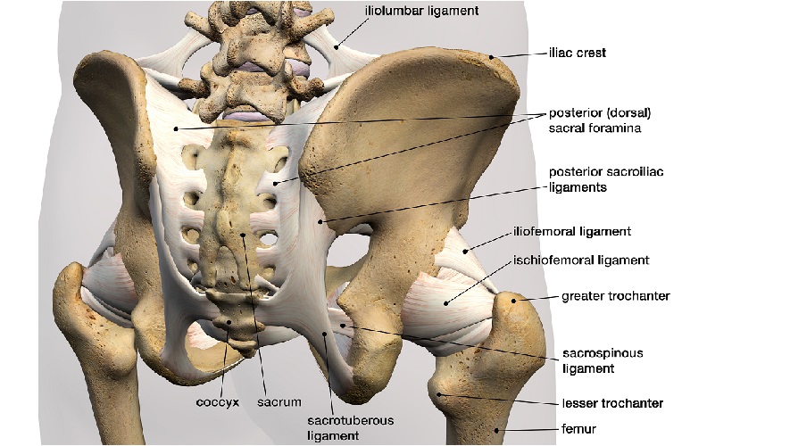
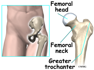
Hip muscle anatomy support movement. Bones of the leg and foot. The two bones beneath your knee that make up your shin are. More than 99 percent of our body's calcium is held in our bones and teeth. The head of your femur fits into your hip socket and the bottom end connects to your knee. Osteoporosis and hip fracture 4995. Simple bone diagram wiring diagram. Your leg bones are the longest and strongest bones in your body. The ilium bone forms the superior portion of the os coxa, the ischium bone the lower posterior portion, and the pubic bone (pubis) the lower anterior portion. These same nerves innervate the knee, which explains why pain can be referred to the knee from the hip and vice versa. Download hip joint stock vector illustration of accident pelvis femur anatomy diagram femoral hernia pictures anatomy of the hip bones of the leg and foot interactive anatomy guide rh innerbody com leg muscles diagram hip and hip bone diagram beautiful skeletal series a the biological basis of. Previously covered was the hip and we shall now cover the femur (upper leg), patella (kneecap) and the tibia and fibula (the two lower leg elements). Want to learn more about it?
The hip joint is one of the most important joints in the human body. Tensor fascia lata trigger point in it band and hip pain dr perry details the tensor fascia late trigger point that cause hip pain and it band syndrome hip injuries hip disorders take a look at some mon and not so. Simple bone diagram wiring diagram. It joins the lower limb to the pelvic girdle. The two bones beneath your knee that make up your shin are.
Lower Extremity Anatomy Bones Muscles Nerves Vessels Kenhub from thumbor.kenhub.com Leg bone diagram julius randle and his future. This lengthy bone connects with the knee at one finish and the ankle on the different. The ilium, ischium, and the pubis. Simple bone diagram wiring diagram. Diagram depicting the arterial supply to a growing leg. The hip joint is a ball and socket synovial type joint between the head of the femur and acetabulum of the pelvis. Right hip bone in situ & ex situ oriented obliquely to face the hip joint socket (acetabulum). The hip joint is one of the most important joints in the human body.
Hip muscle anatomy support movement.
Hip anatomy pictures function problems treatment. The main reason for its in depth use is that they are corrosion resistant and can be employed in any type of surroundings. The bone surfaces of the femoral head and acetabulum have a smooth durable layer of articular cartilage that cushions the ends of the bones and allows for smooth movement. Hip bone femur stock photo image of pelvic articular. Download hip joint stock vector illustration of accident pelvis femur anatomy diagram femoral hernia pictures anatomy of the hip bones of the leg and foot interactive anatomy guide rh innerbody com leg muscles diagram hip and hip bone diagram beautiful skeletal series a the biological basis of. Bones of the leg and foot. Leg bone diagram julius randle and his future. Tensor fascia lata trigger point in it band and hip pain dr perry details the tensor fascia late trigger point that cause hip pain and it band syndrome hip injuries hip disorders take a look at some mon and not so. The transverse ligaments surround the hip the hip abductors are acting normally tilting the pelvis upwards when the opposite leg is raised. Leg bones diagram femur manual e books. Muscles of hip, thigh, leg, and foot. Femur bone diagram, picture of femur bone diagram. The knee is a strong but flexible hinge joint that uses muscles and.
Skeletal hand diagram just another wiring diagram blog. Femur bone diagram, picture of femur bone diagram. Hip anatomy pictures function problems treatment. Click and start learning now! Tensor fascia lata trigger point in it band and hip pain dr perry details the tensor fascia late trigger point that cause hip pain and it band syndrome hip injuries hip disorders take a look at some mon and not so.
Hip Anatomy from www.eorthopod.com Download this free vector about diagram showing the hip bone treatment, and discover more than 13 million professional graphic resources on freepik. The foot bones shown in this diagram are the talus, navicular, cuneiform, cuboid, metatarsals and calcaneus. Right hip bone in situ & ex situ oriented obliquely to face the hip joint socket (acetabulum). Bones are mostly made of the protein collagen , which forms a soft framework. The hip bone (os coxae, innominate bone, pelvic bone or coxal bone) is a large irregular bone, constricted in the center and expanded above and below. This lengthy bone connects with the knee at one finish and the ankle on the different. The knee is a strong but flexible hinge joint that uses muscles and. Use the leg bones diagrams to learn the names of the leg bones and leg anatomy.
Previously covered was the hip and we shall now cover the femur (upper leg), patella (kneecap) and the tibia and fibula (the two lower leg elements).
Simple bone diagram wiring diagram. The femur is the upper leg bone or thigh. Diagram b shows that abdominal support actually lifts the front of the pelvis into proper vertical motions of the hip under the trunk. Bones are mostly made of the protein collagen , which forms a soft framework. The foot bones shown in this diagram are the talus, navicular, cuneiform, cuboid, metatarsals and calcaneus. Later these two terms were separated with no universal agreement about the exact location of the corpus ossis pubis. Bones of the leg and foot. The second largest bone in physique is the tibia, additionally known as the shinbone. Hip muscle anatomy support movement. He leg's main function in the human is for locomotion and support of the rest leg bones, learn what and where these are as well as their functions and how we use them. The knee is a strong but flexible hinge joint that uses muscles and. The hip joint gives the leg an incredible range of motion while still providing support to the body's weight. Leg bones diagram femur manual e books.
Osteoporosis and hip fracture 4995 leg bone diagram. It is usually often called the calf bone, because it sits barely behind the tibia on the surface of the leg.


0 Komentar